The Effective Surface: A Strategy to Unveil the Surface of Proteins

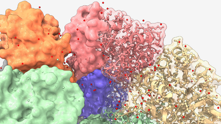
The surface of proteins is irregular, diverse, and exposed to environmental changes, which makes its study challenging. At ICMAB, researchers propose the “effective surface”: a way to measure the protein’s behavior using a small anionic molecule that enables surface mapping. With the help of computational techniques, this promising tool could allow the visualization of these surfaces in great detail and high resolution.
The shape of a protein is not a mere whim: it is determined by the order of its amino acids, by the internal forces that hold it together (such as hydrogen or dihydrogen bonds), and, above all, by its three-dimensional structure, which depends on the environment in which it is found. This shape is key to its function, as it allows the protein to interact with other molecules, as occurs in vital processes within the body.
One of the most important—and at the same time most difficult to study-aspects is its surface. Although we often imagine it as smooth and uniform, the reality is quite different: the surface of a protein is irregular, with complex and wavy forms. Moreover, this surface can change depending on the environment, such as the ions and solvent molecules surrounding it, which makes its study even more challenging.
Traditionally, visualizing a protein in its natural state, without modifying it, has been a major challenge. In our study, we propose a new way of observing it: through what we call its “effective surface”, that is, how it truly behaves when interacting with other molecules, as it would when binding to receptors on a cell membrane.
To achieve this, we use a small anionic molecule called [o-COSAN]⁻, which is very stable and does not alter the protein’s structure. This molecule acts as a kind of “probe” that binds to specific parts of the protein and allows us to map its surface using simple electrochemical measurements, without the need for invasive techniques.
Thanks to this strategy, we can identify which areas of the protein are more reactive or more likely to interact with other substances. This information is extremely valuable for many applications, from the development of new drugs to advances in biotechnology.
What’s most interesting is that [o-COSAN]⁻ “sees” each protein differently, which allows us to classify them based on the unique characteristics of their surface. Thus, this molecule becomes a very useful tool for studying proteins in their natural state.
Although this work has been carried out in the laboratory, we are convinced that, with the help of appropriate algorithms and computational models, we could eventually visualize these surfaces in great detail and high resolution. This would open new doors to scientific research and the design of more precise therapies.
Figure 1. a) Schematic representation of the rotamer cis-[o-COSAN]⁻ with the numbering of its vertices and its ability to form nanovesicles at low concentrations of the rotamer in aqueous solution. b) Two distinct types of protein interactions modeled based on the basic amino acid residues present on the surface of the protein, using the anionic "small molecule" probe: [o-COSAN]⁻.
Institut de Ciència de Materials de Barcelona (ICMAB-CSIC)
UAB Sphere
References
Xavier, J. A. M.; Fuentes, I.; Nuez-Martínez, M.; Viñas, C. & Teixidor, F. (2023). Single stop analysis of a protein surface using molecular probe electrochemistry. Journal of Materials Chemistry B, 11, 8422-8432. http://doi.org/10.1039/d3tb00816a

