Medicine and Health
Pancreas: ultrasounds for a better diagnosis
Non invasive techniques are essential for diagnosis in medicine. One of these techniques is the ultrasonography, an exploration technique that registers the echoes of acoustic waves inside the body. One of the applications of the ultrasounds is the exploration of the pancreas.
References
"Ultrasonography of the pancreas. 1. Conventional imaging". Martínez-Noguera A, D'Onofrio M. Abdom Imaging. 2007 Mar-Apr;32(2):136-49.
The ultrasounds (US)image of the pancreas has meant a big advance in the last years and plays an important role in the detection, characterization and the staging of the pancreatic illnesses. Conventional ultrasonography (US) is a non-invasive imaging modality, which continues to be the first diagnostic step in the evaluation of the pancreas.
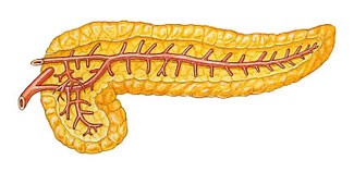
The pancreas is usually divided into head, neck, body, and tail regions. In accordance with the literature, the pancreas can be visualized by US in a big percentage of patients (93 %). To obtain a complete visualization of all portions of the gland is convenient to perform different scanning techniques, such as filling the stomach with water, examining the patient with suspended inspiration or expiration and changing the patient’s position.
In the pathological processes the aptitude to reflect the ultrasounds of the pancreas (its echogenicity) is altered. Among these processes we find acute pancreatitis and its complications, chronic pancreatitis or the pancreatic adenocarcinomas. Using ultrasonography all of these complications can be visualized, acute fluid collections, pseudocyst, pancreatic abscess, infected necrosis and haemorrhage, pseudoaneurysms and venous thrombosis
The clinical diagnosis of chronic pancreatitis is best confirmed together US, CT, magnetic resonance MRcolagiopancreatography (MRCP) and endoscopic retrograde colangiopancreatography (ERCP). Recent technological developments in the field of US, as harmonics and US pulse inversion, color-power Doppler and new contrasts agents allow pancreatic US imaging compete with CT and MR.
Antonio Martínez Noguera
Mirko D’Onofrio2
Department of Medicine
Universitat Autònoma de Barcelona
2 Università di Verona2026 Universitat Autònoma de Barcelona
B.11870-2012 ISSN: 2014-6388