New structural possibilities for the condensation of chromosomes

We have performed a very extensive electron microscopy investigation of the chromatin structures extruded from partially denatured metaphase chromosomes from HeLa cells under a wide variety of conditions. Denatured chromosomes having fibers as the dominant structural element are obtained in the presence of buffers of very low concentration or after incubation with water. At slightly higher ionic concentrations, metaphase chromosomes become granulated. The most frequently observed granules have a diameter of about 35 nm and show the same structural characteristics as the compact cylindrical chromatin bodies previously found in our laboratory in studies performed using small chromatin fragments.
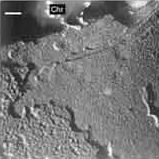
Our results suggest that fibers are formed by the face-to-face association of 35-nm chromatin bodies. We have observed a very compact morphology of chromosomes in solutions containing intracellular concentrations of monovalent cations and the Mg2+ concentration found in metaphase. The most abundant structural elements observed in chromatin extruded from partially denatured compact metaphase chromosomes are multilayered plate-like structures.
This is the first time that these planar structures have been reported. The observation of the irregular plates found in some preparations and of the small planar structures seen in aggregates of small chromatin fragments suggests that plates are formed by side-by-side association of compact chromatin bodies.
Juan Manuel Caravaca, Silvia Caño, Isaac Gállego, Joan-Ramon Daban
References
Article: J.M. Caravaca, S. Caño, I. Gállego i J.R. Daban (2005) "Structural elements of bulk chromatin within metaphase chromosomes" Chromosome Research, 13: 725-743.

