

New model developed to study MERS-coronavirus infection in the laboratory
Researchers at the IRTA-UAB Joint Research Unit in Animal Health have developed and validated a technique for the culture of llama respiratory tissue that permits to study why camelids overcome infection caused by MERS-CoV without developing clinical signs. The methodology will allow to test therapeutic products for humans against this coronavirus, thus reducing the use of animals for experimental purposes.
Middle East respiratory syndrome coronavirus (MERS-CoV) infection causes severe pneumonia, which can be fatal, characterized by a massive infiltration of inflammatory cells into the lungs. Bats and camelids are the natural reservoirs of MERS-CoV. Dromedary camels are the most important vehicles for MERS-CoV evolution and the main source of human infection. The main reasons for these facts include: 1) dromedaries do not experience disease after infection with MERS-CoV, which abundantly replicates in the upper respiratory tract of these animals, 2) individuals are rapidly re-infected in the field, and 3) there is a high risk of infection transmission to camel herders in close contact.
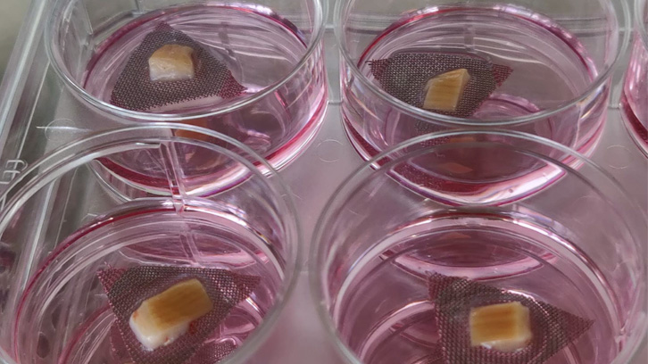
Understanding how camelids clear MERS-CoV infection without displaying clinical signs would aid in the development of human therapies against the virus. Nonetheless, such investigations imply using animals for research purposes, which raises ethical and moral concerns. Therefore, to reduce the number of animals used in experimental research, a model for studying infection and replication of MERS-CoV in the lab has been developed. Using an ex vivo culture of camelid (llama) respiratory tissue, we determined the appropriate viral dose to reproduce the infection in tracheal explants in a similar way that it occurs in vivo. The new model will allow testing of therapeutic products against MERS-CoV while reducing the use of experimental animals.
Furthermore, the study of immune responses in respiratory explants infected with MERS-CoV leads to similar results in experimentally inoculated animals. Our research group previously published a study demonstrating that the infected nasal epithelium of camelids activates essential defence molecules, known as interferons (IFNs), which activate antiviral immune responses (IFN-stimulated genes or ISGs) along the respiratory tract to clear viral infection (Te et al., 2021). Using the novel alternative infection model, we confirmed that IFNs are not originated at the tracheal level. This study supports the hypothesis that ISGs expression in the trachea of experimentally inoculated camelids is a direct consequence of IFN production at the nasal cavity and the interaction between both tissues.
1) Joint Research Unit IRTA-UAB in Animal Health, Centre de Recerca en Sanitat Animal (CReSA)
2) IRTA, Animal Health Programm, Centre de Recerca en Sanitat Animal (CReSA
3) School of Public Health, Li Ka Shing, Faculty of Medicine, The University of Hong Kong
4) Departament of Animal Health and Anatomy, Universitat Autònoma de Barcelona
References
Te, N., Rodon, J., Creve, R. et al. Evaluation of alpaca tracheal explants as an ex vivo model for the study of Middle East respiratory syndrome coronavirus (MERS-CoV) infection. Vet Res 53, 67 (2022). https://doi.org/10.1186/s13567-022-01084-3