Key biomarkers of tumorigenesis and malignancy identified in canine mammary neoplasia
A study conducted by the UAB has identified potential biomarkers of tumorigenesis and malignancy in canine mammary neoplasia (CMN). The researchers have analyzed gene expression in tumor and healthy mammary tissue samples using the molecular technique qRT-PCR. These findings could improve early diagnosis and treatment of affected canines.
This work was carried out in close collaboration between the Faculty of Veterinary Medicine and the Department of Cell Biology, Physiology, and Immunology both from Universitat Autònoma de Barcelona, as part of the work of a recently presented PhD thesis. The authors searched for new potential molecular markers of malignancy using gene expression in canine mammary neoplasms (CNM).
Early diagnosis of tumors, especially malignant ones, is crucial in current cancer therapy strategies to improve the prognosis. Nowadays, the study of tumor-linked gene expression patterns has gained relevance as a useful tool in the diagnosis, prognosis, and treatment of CMN. Several biomarkers have been widely evaluated in breast cancer, but research on CMN is less abundant.
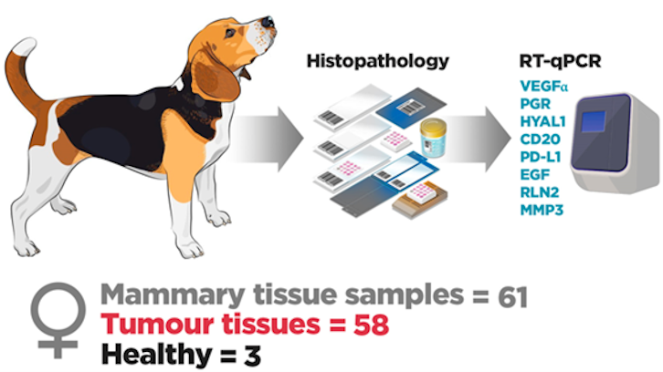
For this reason, we conducted a pilot study to assess the gene expression of hyaluronidase-1 (HYAL-1), CD20, matrix metallopeptidase 3 (MMP3), vascular endothelial growth factor-α (VEGFα), relaxin 2 (RLN2), programmed death-ligand 1 (PD-L1), epidermal growth factor (EGF), and progesterone receptor (PGR) in CNM to determine their putative potential as molecular biomarkers of malignancy through the molecular technique qRT-PCR. Fifty-eight formalin-fixed paraffin-embedded (FFPE) paraffined CMN were included and histologically classified as benign (n=37) or malignant (n=31). In addition, mammary samples from three healthy bitches were also included.
Figure 1: Relative mRNA levels of target genes measured in samples obtained from healthy mammary tissue and mammary tumours. Bars represent the median value with the interquartile range. Statistical significances between groups are marked with asterisks (* p < 0.05; ** p < 0.01; *** p<0.001).
All the assessed genes showed higher expression in neoplastic mammary tissue than in healthy tissue. That is, they were overexpressed in neoplastic mammary tissue, suggesting a role in the process of tumorigenesis (Figure 1). Moreover, PD-L1, EGF, relaxin, and MMP3 were significantly overexpressed in malignant canine mammary neoplasia compared to benign canine mammary neoplasia, suggesting they may be useful as malignancy biomarkers.
Finally, we can conclude that the over-expression of the evaluated genes is related to the tumorigenic process in canine mammary neoplasia, whereas PDL-1, EGF, RLN2, and MMP3, may be also useful as malignancy biomarkers for CMN purposes.
Mariana Teles
Department of Cell Biology, Physiology and Immunology
Universitat Autònoma de Barcelona
References
Galadima, M.; Teles, M.; Pastor, J.; Hernández-Losa, J.; Rodríguez-Gil, J.E.; Rivera del Alamo, M.M. Programmed Death-Ligand (PD-L1), Epidermal Growth Factor (EGF), Relaxin, and Matrix Metalloproteinase-3 (MMP3): Potential Biomarkers of Malignancy in Canine Mammary Neoplasia. Int. J. Mol. Sci. 2024, 25, 1170. https://doi.org/10.3390/ijms25021170